Figure 7.

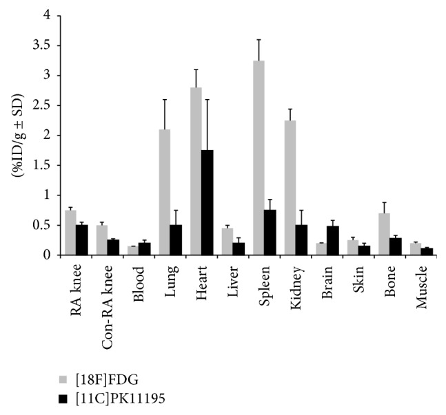
Ex vivo tissue distribution of [18F]FDG (n = 6), light grey bars, and (R)-[11C]PK11195 (n = 5, black bars) at 1 h after injection in rats of group A (6 d). Results are expressed as percentage of the injected dose per gram (%ID/g ± SD).

Ex vivo tissue distribution of [18F]FDG (n = 6), light grey bars, and (R)-[11C]PK11195 (n = 5, black bars) at 1 h after injection in rats of group A (6 d). Results are expressed as percentage of the injected dose per gram (%ID/g ± SD).