Fig. 2.
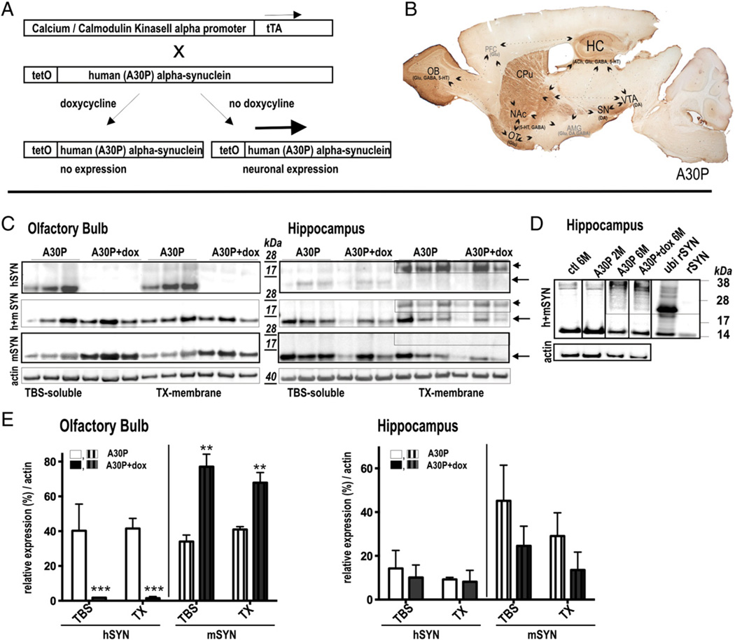
Expression pattern and regulatability of human A30P α-syn in subregions of conditional mouse brain (A) In double transgenic mice, expression of human A30P α-syn gene is under control of the tetracycline-controlled transactivator (tTA) responsive promoter (tetO) and tTA expression under the control of the CaMKIIa promoter, leading to neuron-specific expression. Expression is abolished by tetracycline derivates (doxycycline; dox) as it binds to tTA rendering it incapable of binding to tetO sequence. (B) Sagittal overview showing strong expression of human A30P α-syn in the (OB), the substantia nigra (SN), the ventral tegmental area (VTA), the nucleus accumbens (NAc), the caudate–putamen (CPu), the olfactory tubercle (OT) and the hippocampus (HC). These brain regions are implicated in resilience to anxiety disorders and are reported to regulate stress response [modified from (Russo et al., 2012) with the OB circuitry added in accordance to its function in depression (Song and Leonard, 2005)]. (C) Immunoblots of olfactory and hippocampal brain extracts using antibodies against human α-syn (hSYN), mouse α-syn (mSYN) and mouse and human α-syn (h + mSYN). Ultracentrifugation and sequential extraction steps were used to obtain TBS-sol-uble and TX-membrane fractions of α-syn in A30P mice with and without treatment of dox. Tight regulation of 14 kDa soluble and membrane bound α-syn was detected in the OB whereas in the HC ∼25 kDa dox-resistant α-syn species accumulated in the membrane fraction. Note that in the OB the endogenous α-syn was opposingly expressed. (D) HC α-syn and ubiquitinated recombinant α-syn expression pattern. (E) Quantification of human and murine α-syn expression levels (n = 3 per group). Results are expressed as mean + SEM. **p < 0.01; ***p < 0.001 (two-tailed t-test).

