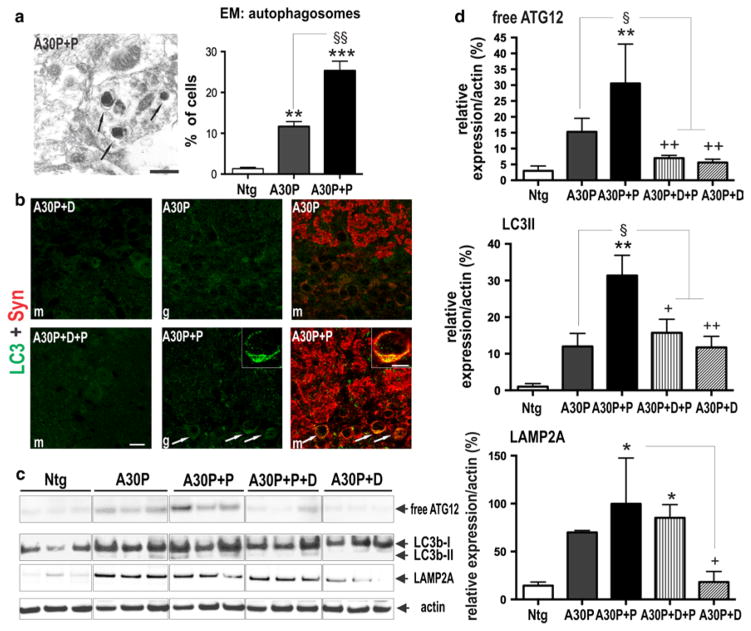
Fig. 7.
Reduced autophagolysosome clearance in OB of A30P+P mice. a Representative electron micrographs of GL from A30P+P mice. Clustered autophagosomal structures with higher electron density were increased in A30P+P mice. Right number of autophagosomes per cell profile (50 profiles per group). b Double-fluorescence confocal photomicrographs showed a relative strong accumulation of LC3 immunoreactive puncta colocalizing with human α-syn in A30P+P mice (see also inset) when compared to A30P or dox-treated controls. c Protein extracts from OB and striatum increase detected with antibodies against membrane proteins of the autolysosme. d Quantification revealed increase in free ATG12, and autolysosome membrane marker Lamp2A and reduced clearance of autolysosome substrate LC3-II in OB neurons of A30P+P mice. Immunoblots are highlighted by separating boxes for clarity of display. The data are expressed as mean ± SEM. **p < 0.01 vs. Ntg, §p < 0.05 vs. A30P, +p < 0.05, ++p < 0.05, vs. A30P+D. Scale bar 1 μm in a, 10 μm in b and 5 μm in inset

