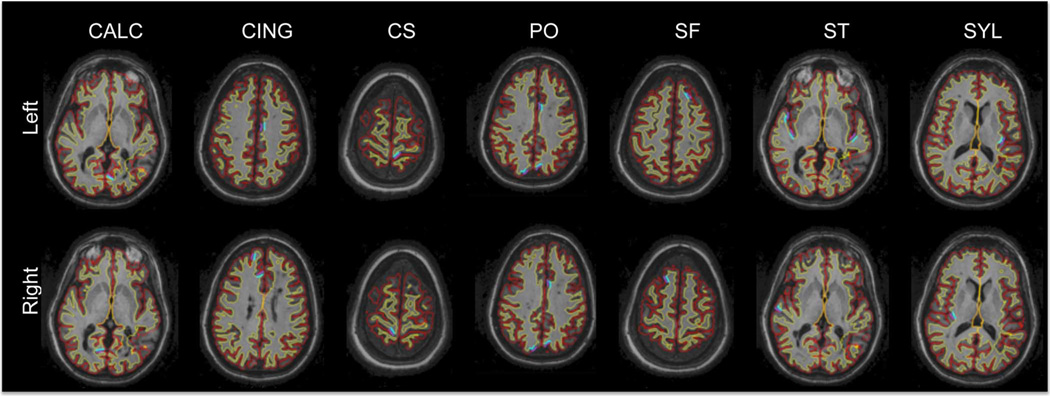
Fig. 8.
Outline of the reconstructed FreeSurfer surfaces on a series of horizontal slices for an MS patient. Red: GM surface, yellow: WM surface. Manually picked landmark clusters are shown in purple (GM) and blue (WM). CALC: Calcarine sulcus, CING: cingulate, CS: central sulcus, PO: parieto-occipital sulcus, SF: superior-frontal sulcus, ST: superior-temporal sulcus, SYL: Sylvian fissure.

