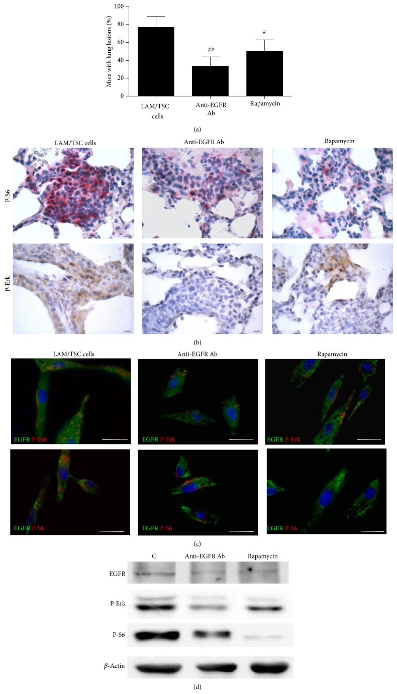
Figure 2.
Anti-EGFR antibody (Ab) reduces lung nodules caused by LAM/TSC cell administration and phosphorylation of S6 and Erk. (a) Anti-EGFR Ab (n = 13) reduces the percentage of mice with lung nodules 30 weeks after cell administration more efficiently than rapamycin (n = 11). Data are means ± SEM. # P < 0.05; ## P < 0.01 versus mice 30 weeks after cell administration (n = 13). (b) Immunohistochemical analysis with phosphorylated S6 antibody at Ser235/236 (red staining, arrows) and phospho-Erk antibody at Thr202/Tyr204 (brown staining) in lung nodules 30 weeks after cell administration and after anti-EGFR antibody and rapamycin treatment. Scale bars: 10 μm. (c) The expressions of EGFR (green labelling) and P-Erk (red labelling, upper panels) or P-S6 (red labelling, lower panels) are detected in LAM/TSC cells, 24 hours after anti-EGFR Ab or rapamycin incubation. Scale bars: 50 μm. (d) Western blotting analysis shows the decreased levels of P-S6 and P-Erk following anti-EGFR Ab and rapamycin for 24-hour incubation in LAM/TSC cells. β-Actin is used as loading control.

