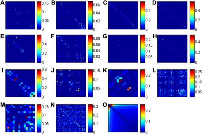
Fig 1. Coevolution patterns in 15 HIV-1 proteins.

The panels (A-L) are heat maps of the direct information (DI) values of residue pairs in multiple sequence alignments of GP120, GP41, MA, CA, NC, PR, RT, IN, P6, NEF, REV, TAT, VIF, VPR, and VPU, respectively. The x- and y-axes represent the positions of amino acid residues in the multiple sequence alignments with gap filtering (see Methods).
