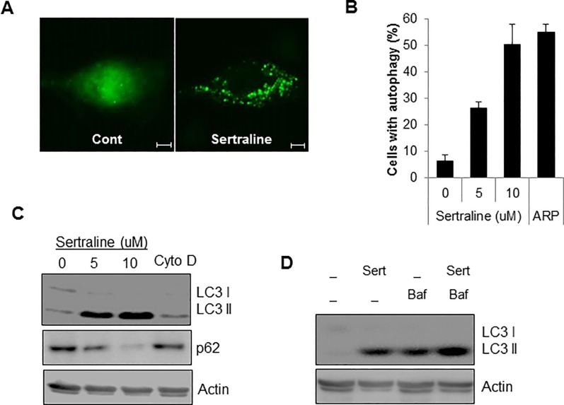
Fig 3. Sertraline activates autophagy in htRPE cells.
(A) htRPE/GFP-LC3 cells were treated with sertraline (10 uM) and fixed to for the fluorescence imaging (Scale bars: 20 μm). (B and C) htRPE/GFP-LC3 cells were exposed to either increasing concentration of sertraline (5, 10 μM) or cytochalasin D (Cyto D, 50 nM) for 24 h. Cells with autophagic punctuate structures were counted (B). Cells were harvested to analyze Western blotting with indicated antibodies (C). (D) htRPE cells were treated with Sertraline (Ser, 10 μM) with or without an autophagy inhibitor, bafilomycin A1 (Baf). The expression level of LC3 protein was detected by Western blotting. Data were obtained from at least three independent experiments and values are presented as the means ± S.E.M. (n > 3, * p < 0.02).

