
Figure 2.
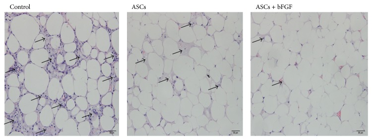
HE stain evaluation of fat grafts (magnification ×200). The control grafts exhibited excessive fibrosis (shown as ↑); in the group with ASCs, the grafts consisted predominantly of mature lipocyte and had significant lower levels of fibrosis; and bFGF ameliorated fibrosis more.