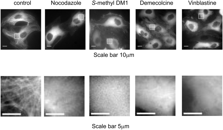
Fig 3. Fluorescent microscopy photographs of ECFP-PTK2 cells treated with various microtubule depolymerizing agents.
Each compound was added into culture medium with the following final concentration and incubated for the following time period: nocodazole (0.3 μM for 60 min), S-methyl DM1 (0.2 μM for 90 min), demecolcine (1.6 μM for 90 min) and vinblastine (1 μM for 90 min). Scale bars are 10 μm for main images and 5 μm for sub-region images.

