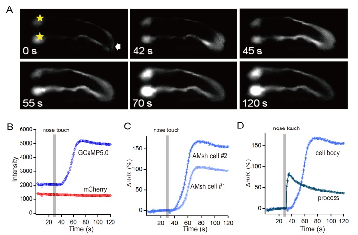
Fig 3. In vivo tactile stimulation induced Ca2+ elevation in C. elegans AMsh cell.
(A) An example of intracellular Ca2+ elevation in an AMsh cell induced by a train of tactile stimuli (20 μm, 2 Hz, 5 s). Sample times are indicated in seconds. *, cell body of the AMsh cell. (B) Fluorescence intensities in the cell body of an AMsh cell. (C) Ratio changes in the cell bodies of the AMsh cells. (D) Ratio changes in the process and cell body of an AMsh cell. The Ca2+ wave was propagated from the process to the cell body.

