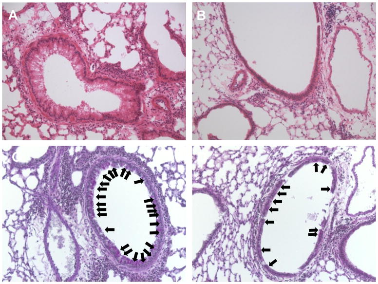
Figure 2. Decreased airway inflammation and mucus metaplasia in Fat-1 transgenic mice.
Lung tissue from WT (left) and Fat-1 (right) mice was obtained and prepared for histopathological analysis (see Methods). (A,B) Lung sections stained with H & E revealed decreased leukocyte infiltration and less reactive airway epithelium in the lungs of Fat-1 mice compared to WT littermate controls. (C,D) Periodic Acid-Schiff staining revealed decreased mucus (arrows) in the airway epithelium in Fat-1 mice as compared to WT controls. Images are representative for n = 3 in each cohort.

