
FIG 3.
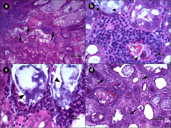
Microphotographs of hepatopancreases (HPs) from L. vannamei naturally infected by AHPND in the terminal stage of infection. (a) Transversal view of HP with severe hemocytic infiltration in the interstitial spaces as a response to epithelium necrosis (arrows). (b and c) Hepatopancreas tubule shows hemocytic infiltration, with necrotic cells (asterisk) and bacterial masses inside the lumen (arrowheads). (d) Transverse view of HP with severe hemocytic infiltration (arrow) and melanization of necrotic material inside the tubules. Hematoxylin and eosin staining was used. Microphotographs by S. Abad.