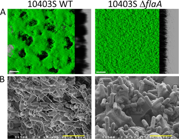
FIG 6.

Microscopic observations of the biofilms formed by the motile L. monocytogenes 10403S WT strain and its isogenic nonmotile 10403S ΔflaA mutant. (A) Isosurface representation obtained from the confocal image series using the IMARIS software (green Syto 9 staining). (B) SEM image at 1.5 × 10⁴ magnification. The white scale bars correspond to 30 μm and the yellow bars to 2 μm.
