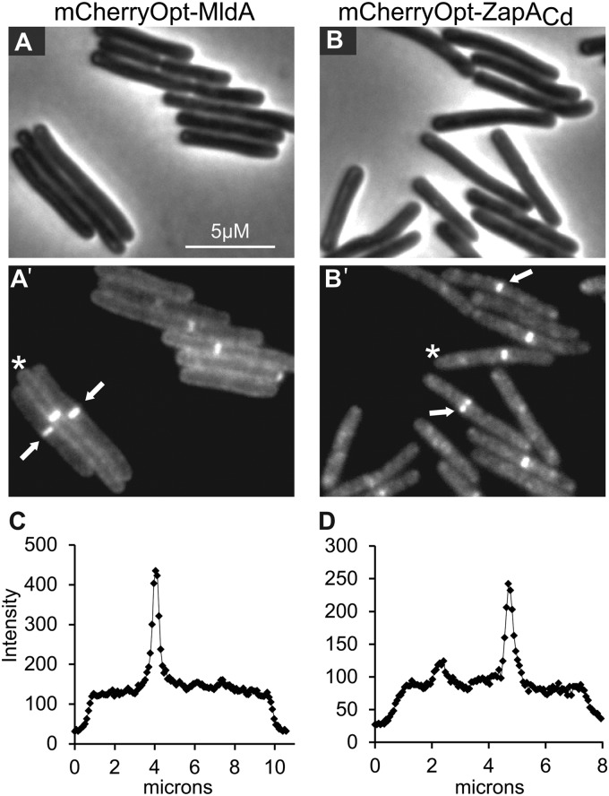
FIG 4.
Septal localization of mCherryOpt fusions to the division proteins MldA and ZapA. C. difficile cells harboring pRAN535 (mCherryOpt-MldA) or pRAN534 (mCherryOpt-ZapACd) were fixed, removed from the anaerobic chamber, and photographed the next morning. (A and B) Phase-contrast images. (A′ and B′) Fluorescence images. Arrows indicate examples of septal localization. (C and D) Quantitation of fluorescence intensity along transects through the cells marked with an asterisk in panels A′ and B′. The transects begin ∼1 μm before each cell (i.e., the end with the asterisk) and extend ∼1 μm beyond each cell. As noted elsewhere (6) and as documented in Fig. S3 in the supplemental material, C. difficile does not always divide precisely at the midcell.

