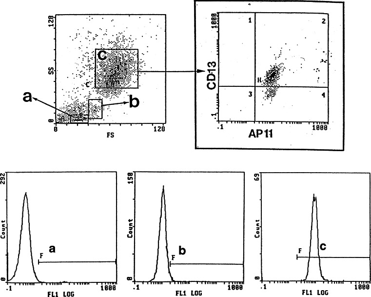
Fig. 1.
In-flow cytometric analysis of peripheral blood using mAb AP11. Granulocytes show a high level of reactivity to mAb AP11. Granulocytes (c) were gated to determine the CD13 and AP11 positivity (upper right panel). Lower panels show fluorescence for AP11 for lymphocytes (a), monocytes (b), and granulocytes, respectively

