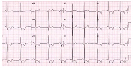
Figure 6.

Patient 6. An electrocardiographic (ECG)tracing, showing voltage criteria for left ventricular hypertrophy with ST depression and negative T wave in the precordial and inferior leads V3-6, of a 52-year female patient with non-obstructive hypertrophic cardiomyopathy (thickness of septum 20 mm and posterior wall of 24 mm without septal anterior movement or obstruction of outflow tract). Normal coronary arteries were found on coronary angiography. She refused genetic counseling and invasive intervention. She was treated medically with beta blocker.
