
Figure 8. SCC categorized as NSCLC analyzed by in situ hybridization for miR-199a-5p or immunohistochemistry for Brm, CD44, MET, CAV1.
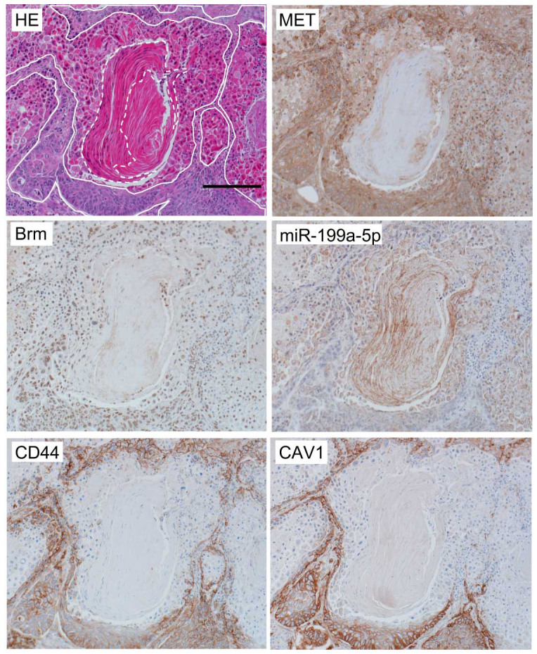
The bar indicates 200 μm. In the HE staining slide, less differentiated cells are surrounded by white solid lines and highly differentiated cells which still retain cellular nuclei at the periphery of a cancer pearl are shown by a white broken line, respectively.