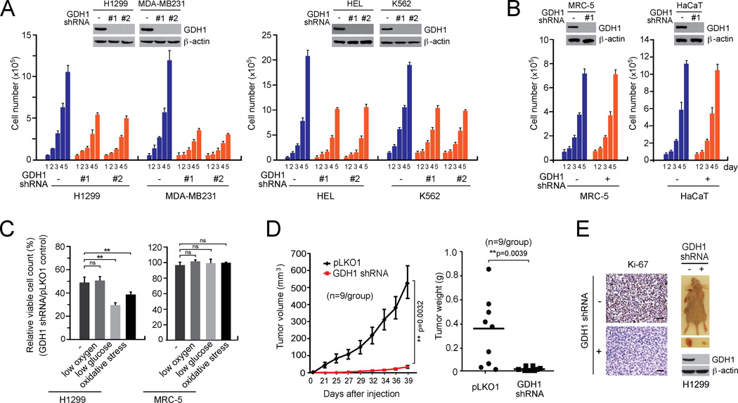
Figure 2. GDH1 is important for cancer cell proliferation and tumor growth.
(A–B) Cell proliferation rates were determined by cell counting in H1299 and MDA-MB231 tumor cells (A; left), HEL and K562 leukemia cells (A; right) and MRC-5 and HaCaT (B) with stable knockdown of GDH1, compared to control cells expressing an empty vector. Expression of GDH1 in cells transduced with GDH1 shRNA clones are shown by Western blot analyses. (C) Effect of GDH1 knockdown on cell proliferation rates were measured under stress conditions including low oxygen (1% O2), low glucose (0.5 mM glucose) and oxidative stress (15 µM H2O2). (D) Effect of GDH1 knockdown on tumor growth potential of H1299 cell xenograft mice. Left: Tumor size was monitored every 2–3 days for 6 weeks. The error bars represent SEM. Right: Tumor weights were examined at the experimental endpoint. (E) Left: Representative pictures of IHC staining to detect Ki-67 expression in tumors harvested from vector control group or GDH1 knockdown group. Scale bars = 50 µm. Right: Representative dissected tumors and GDH1 expression in tumor lysates are shown. Data are mean ± SD from three replicates of each sample except panels D and E. p values were determined by a twotailed Student’s t test for panel C and a two-tailed paired Student’s t test for panel D (ns: not significant; **: 0.001 < p < 0.01). See also Figure S1.

