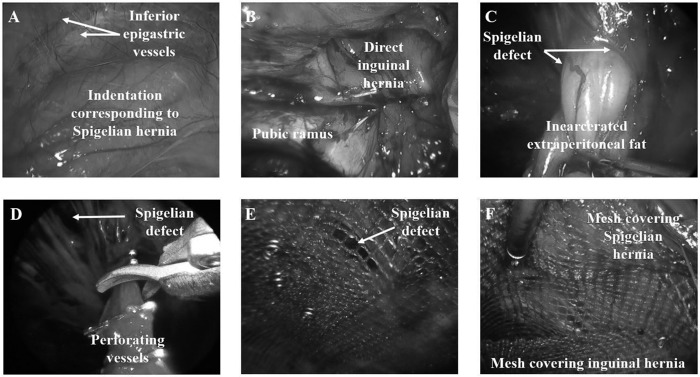
Figure 2.

Intraoperative views of a patient (from Figure 1) presenting with right Spigelian and direct inguinal hernia undergoing SIL Spigelian and inguinal hernia repair with mesh. (A) Intraperitoneal view of site of Spigelian hernia. (B) Direct inguinal hernia. (C) Incarcerated extraperitoneal fat via sharp small defect in the transversus abdominis. (D) Perforating blood vessels being clipped and divided to achieve adequate proximal clearance for mesh placement. (E) Mesh covering Spigelian defect. (F) Mesh covering the inguinal hernia to cover the inferior aspect of mesh covering Spigelian defect.
