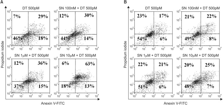
Fig. 3.
Apoptosis after combined treatment with docetaxel at 500pM and selenium at 100nM, 1µM, or 10µM in the MDA-MB-231 cell line (A) and the MCF-7 cell line (B). All cells were stained with fluorescein isothiocyanate (FITC) conjugated Annexin V in a buffer containing propidium iodide and were analyzed using flow cytometry. For each treatment, the percentage of viable cells is shown in the lower left quadrant, for which both Annexin V and propidium iodide levels are low. Data from a representative experiment (from a total of three) are shown.

