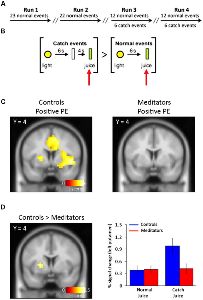
FIGURE 1.
Task outline and positive PE signals. (A) Outline of the conditioning task. fMRI scanning consisted of four separate sessions/runs. Catch events were interspersed among the normal events in run 3 and run 4. Run 1 and run 2 consisted on normal (training) trials only. (B) Normal events consisted of a yellow light (1 s) predicting the oral delivery of fruit juice (0.8 ml) 6 s later. Catch events designed to capture a positive reward PE consisted of presentation of the light cue (1 s) and juice delivery 10 s later at an unexpected time. The specific contrast designed to capture the positive PE was: [Juice delivered (unexpected) > Juice delivered (expected)]. (C) Left panel, positive PE for controls display activity in bilateral putamen. Right panel, positive PEs in meditators did not yield significant voxels in the putamen (see Table 1 for complete list of activations). (D) Left panel, group differences to positive reward PEs show SVC-corrected activity in left putamen in controls. Right panel, parameter estimates for the significant voxels in left putamen show an increase in the BOLD signal at times when juice was not expected but delivered. Controls shown in blue and meditators in red. Error bars indicate SE.

