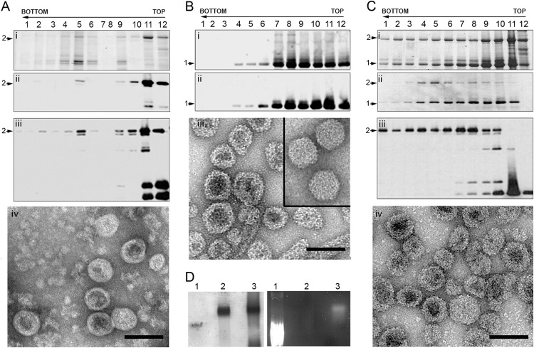
FIG 2.
Expression of the HT-VP2-466-based chimeric VLP and optimization of its assembly. (A) EGFP-HT-VP2-466 chimeric protein was expressed in insect cells, and the assemblies were purified on sucrose gradients; 12 fractions (indicated by the numbers over the lanes) were collected, concentrated, and analyzed by SDS-PAGE and Coomassie staining (i) and by Western blotting using anti-VP2 (ii) and anti-GFP (iii) antibodies. The direction of sedimentation was from right to left, with fraction 12 being at the gradient top. (iv) The image shows a representative electron micrograph (negative staining) of particulate material in fraction 6. Bar = 100 nm. (B) Wild-type HT-VP2-466 (control) was analyzed as described for panel A in a Coomassie-stained gel (i) or by Western blotting for anti-VP2 antibodies (ii). (iii) The image shows HT-VP2-466 assemblies from fraction 6 that correspond to tubes and isometric capsids with a morphology and size similar to those of IBDV capsids (inset). Bar = 100 nm. (Ci to Ciii) Assemblies from cells coexpressing EGFP-HT-VP2-466 and HT-VP2-466 at a 5:1 ratio were analyzed as described for panel Ai to Aiii, respectively. (iv) The electron micrograph shows fraction 6. Bar = 100 nm. (A to C) Arrows labeled 1, HT-VP2-466 (∼50-kDa) bands; arrows labeled 2, EGFP-HT-VP2-466 (∼75-kDa) bands. (D) Native agarose gel electrophoresis after Coomassie staining (left) or UV illumination (right) of EGFP (lanes 1), HT-VP2-466 VLPs (lanes 2), and EGFP-HT-VP2-466 VLPs (lanes 3). Images of the same gel are shown.

