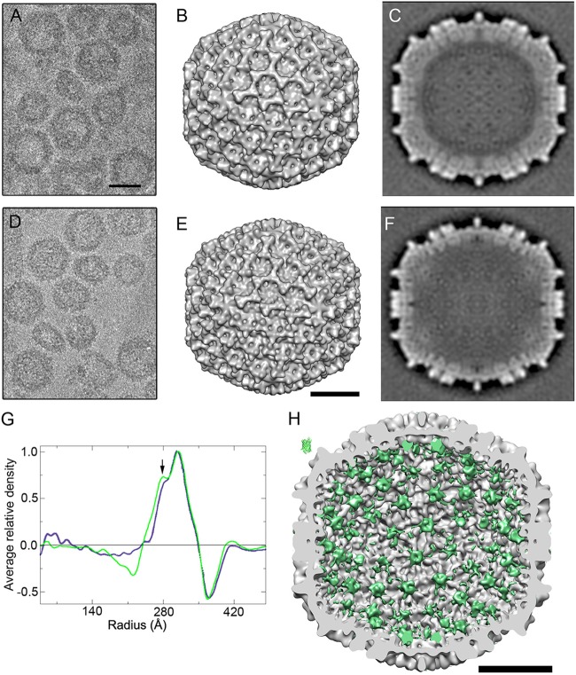
FIG 3.
Three-dimensional cryo-EM reconstructions of chimeric EGFP-HT-VP2-466 and wild-type HT-VP2-466 capsids. (A, D) Cryo-electron micrographs of purified EGFP-HT-VP2-466 (A) and HT-VP2-466 capsids (D). Bar = 50 nm. (B, E) Surface-shaded representations of the outer surface, viewed along an icosahedral 2-fold axis, of the T=13 capsids of EGFP-HT-VP2-466 (B) and HT-VP2-466 (E). Bar = 20 nm. (C, F) Transverse central sections from the 3DR of EGFP-HT-VP2-466 (C) and HT-VP2-466 (F) T=13 VLPs. Lighter shading indicates a higher density. (G) Radial density profiles from 3D maps of EGFP-HT-VP2-466 (green) and HT-VP2-466 (blue) T=13 VLPs computed at a 23-Å resolution. Arrow, extra peak of density on the inner surface of the EGFP-HT-VP2-466 capsid (radius = ∼280 Å). (H) Difference map calculated by arithmetic subtraction of the map for the HT-VP2-466 capsid from the map for the EGFP-HT-VP2-466 capsid. The map on the inner surface of a HT-VP2-466 capsid viewed along an icosahedral 2-fold axis is shown in green. The GFP atomic structure is shown at the same scale (top left). Bar = 20 nm.

