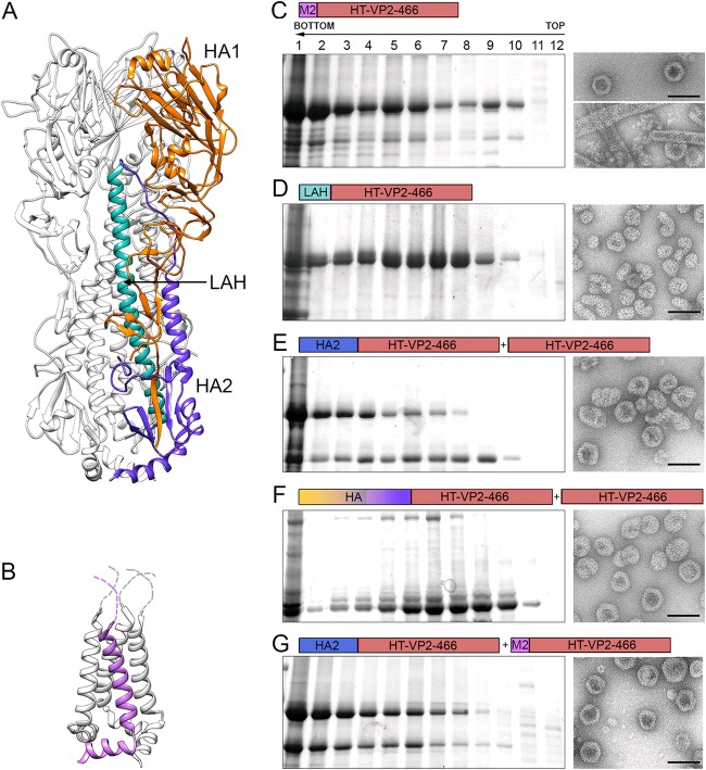
FIG 5.
Expression and purification of influenza virus-derived HT-VP2-466-based chimeric assemblies. (A) Structure of the influenza virus hemagglutinin. Ribbon diagram of an HA trimer (PDB accession number 1RU7), with one monomer colored for clarity (orange and blue, HA1 and HA2 chains, respectively; light blue, LAH). (B) Structure of the influenza virus M2 protein. Tetrameric M2 (PDB accession number 2KWX) is shown as a ribbon diagram, with one monomer shown in violet. The unstructured N-terminal M2 ectodomains are represented as dashed lines. (C to G) Assemblies were purified as described in the legend to Fig. 2A and then analyzed by SDS-PAGE, Coomassie staining, and negative-staining EM. Cells were infected with rBV/M2-CAP (C), rBV/LAH-CAP (D), rBV/HA2-CAP+rBV/CAP (E), rBV/HA-CAP+rBV/CAP (F), or rBV/HA2-CAP+rBV/M2-CAP (G). Electron micrographs (right) in panel C are for fractions 6 (top) and 2 (bottom), and micrographs to the right of panels D to G are for fraction 6. Bars = 100 nm.

