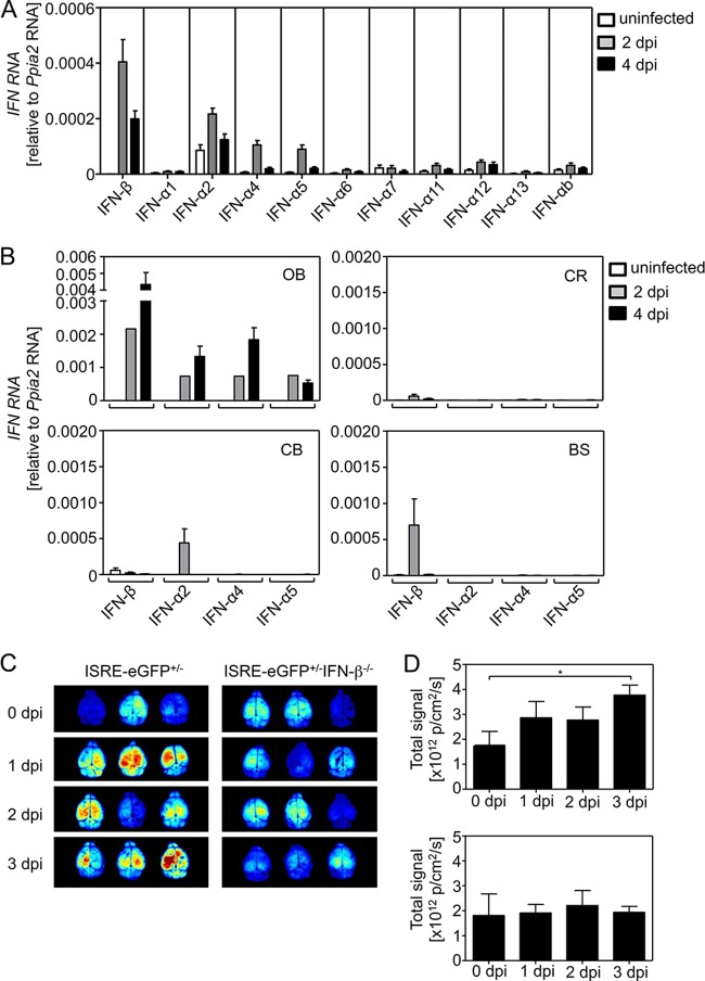
FIG 3.
IFN-β induced locally within the olfactory bulb also stimulates distal parts of the CNS. (A) Mice were i.n. infected with 103 PFU of VSV. At 2 and 4 days p.i. the mice were perfused, and the brains were prepared and lysed in TRIzol. RNA was isolated, purified, and used for cDNA synthesis. To analyze IFN mRNA expression, IFN-subtype-specific TaqMan PCRs were performed. (B) Mice were treated as described above. After perfusion, the brains were prepared and dissected into olfactory bulbs (OB), cerebra (CR), cerebella (CB), and brain stems (BS). Brain regions were lysed in TRIzol, and RNA was isolated. After a purification step, cDNA synthesis and TaqMan PCR were performed for IFN-β, IFN-α2, IFN-α4, and IFN-α5. (C) ISRE-eGFP+/− and ISRE-eGFP+/− IFN-β−/− reporter mice were i.n. infected with 103 PFU of VSV. At the indicated time points, mice were sacrificed, and the eGFP expression was analyzed in a CRI Maestro 2 fluorescence imager (Intas). Representative samples of three independent experiments (15 mice per group in total) are shown. A spectral unmixing algorithm was used to subtract background autofluorescence. (D) The total signal of the brains was evaluated by definition of equal circular regions of interest over the entire brain using the provided software (*, P ≤ 0.05 [Mann-Whitney test]).

