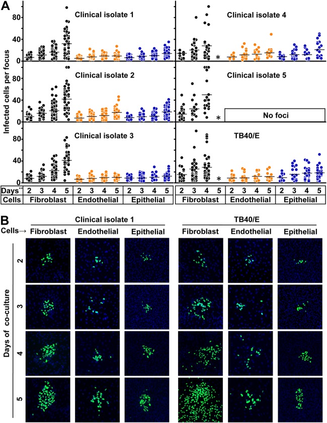
FIG 1.
Kinetics of HCMV transmission in different cell types. (A) Fibroblasts infected by clinical isolates and laboratory strain TB40/E were cocultured with a 2,000-fold excess of uninfected fibroblasts, endothelial cells, or epithelial cells for 2, 3, 4, and 5 days. Monolayers were fixed at the indicated times, and newly infected cells were monitored by HCMV IEA staining. The numbers of infected cells per focus were counted. One dot represents the number of infected cells of an individual focus. Bars indicate mean values of all foci. (B) Representative infectious foci of clinical isolate 1 and TB40/E in fibroblasts, endothelial cells, and epithelial cells are shown. The presence of HCMV IEA (green fluorescence) indicates infected cells, and cell nuclei are stained in blue (DAPI). *, the foci could not be counted due to the high infection rate in fibroblasts; **, days of coculture.

