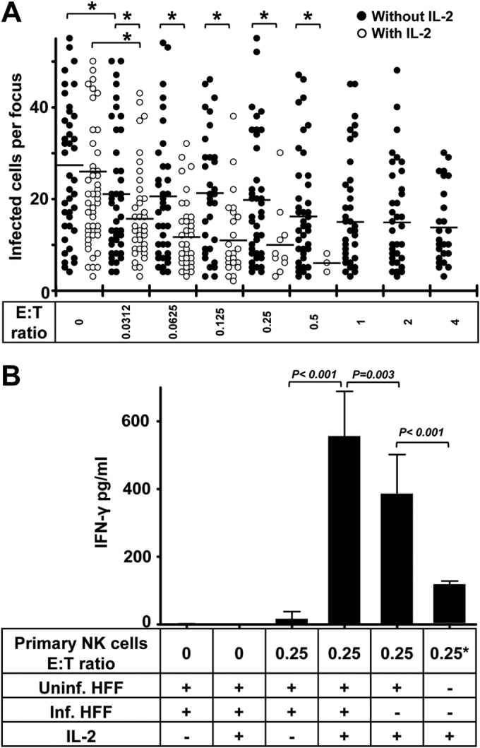
FIG 3.

IL-2 enhances the inhibition of HCMV transmission by primary NK cells. (A) Thawed PBMCs were cultured using NK cell medium with or without IL-2 for 20 h. Next, NK cells were negatively selected from PBMCs. TB40/E-infected fibroblasts were cocultured with a 2,000-fold excess of uninfected fibroblasts for 3 days. Purified NK cells were added to the focus expansion assay immediately at different E:T ratios with or without IL-2-containing medium. Monolayers were fixed at the indicated times, and newly infected cells were monitored by HCMV IEA staining. The numbers of infected cells per focus were counted. One dot represents the number of infected cells of an individual focus. Bars represent mean values of all foci from three donors tested. *, P < 0.05. (B) The concentrations of IFN-γ in supernatants from indicated cultures were tested by ELISA. Uninf., uninfected; Inf., infected. “0.25*” indicates that the same amount of NK cells was used.
