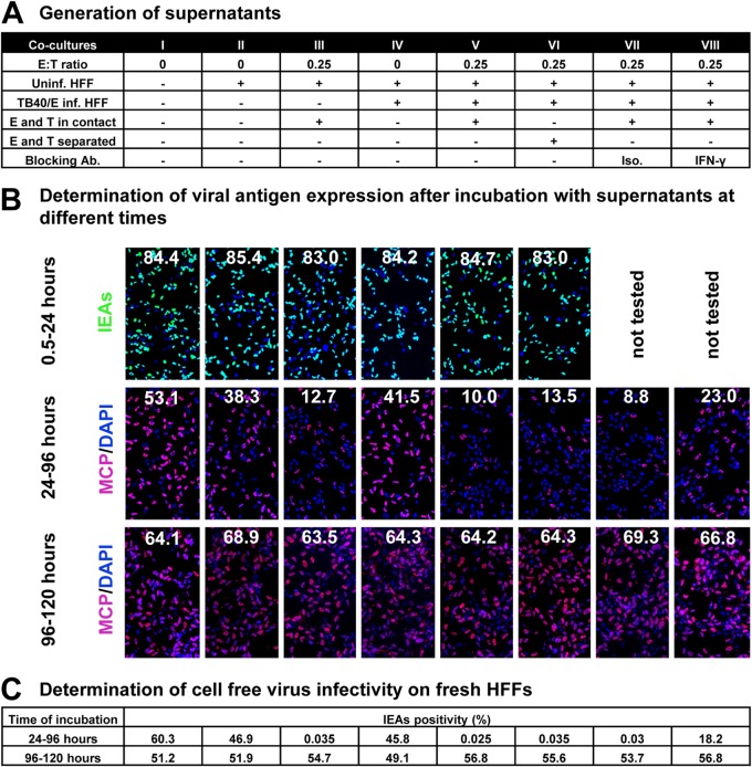
FIG 5.
NK cell-containing supernatants reduce HCMV production. (A) Supernatants were collected from the indicated cocultures and filtered through a 0.1-μm-pore filter to remove cell-free virus. Uninf., uninfected; Inf., infected; Ab, antibody; Iso., isotype antibody control. (B) Fibroblasts were infected by TB40/E (MOI, 7.5) for 30 min and washed with PBS. Thereafter, supernatants were added to infected fibroblasts from 30 min postinfection (p.i.) until 24 h p.i., from 24 to 96 h p.i., or from 96 to 120 h p.i., and then infection rates were determined by HCMV IEA staining. IEA staining is shown by green fluorescence and MCP staining is red at late times infection, and cell nuclei are stained in blue (DAPI). The infection rates (IEA/DAPI) are indicated by the numbers. The MCP-to-DAPI surface area ratios are also indicated by the numbers. (C) Fibroblasts were infected by TB40/E (MOI, 7.5) for 30 min and washed with PBS. Thereafter, supernatants were added to the infected fibroblasts from 24 to 96 h p.i. or from 96 to 120 h p.i. Then 100 μl of 8-fold-diluted supernatants containing cell-free virus was transferred to uninfected fibroblasts. After 30 min of incubation, cells were washed with PBS, and the infection rates (IEA/DAPI) were determined after 24 h.

