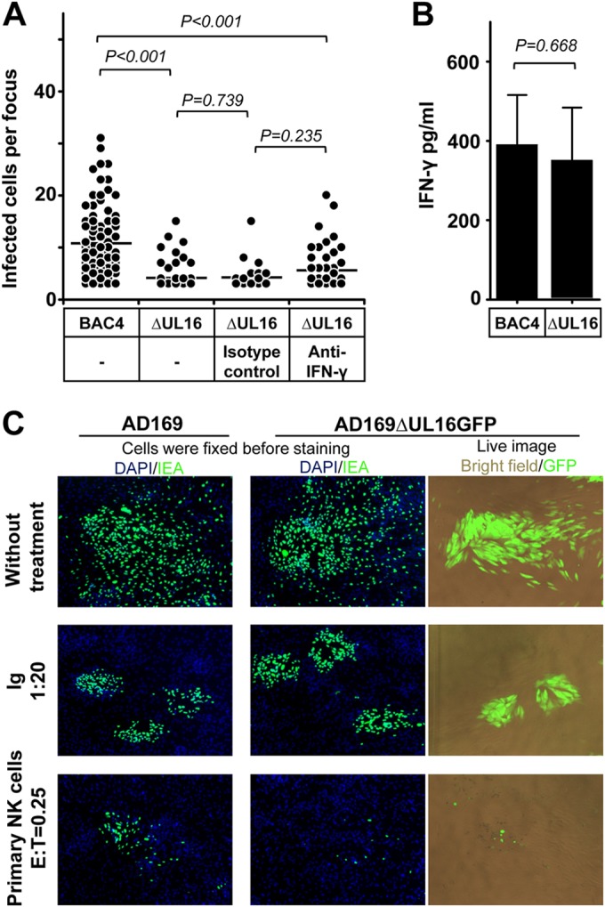
FIG 6.

HCMV gene UL16 modulates the control of HCMV transmission by NK cells. (A) Thawed PBMCs were cultured using NK cell medium with IL-2 for 20 h, and then NK cells were negatively selected from PBMCs. Fibroblasts infected with BAC4 or BAC4ΔUL16 were cocultured with a 2,000-fold excess of uninfected fibroblasts for 3 days. Purified NK cells were added to the focus expansion assay from the beginning at an E:T ratio of 0.25 with IL-2-containing medium. Specific blocking MAb against IFN-γ or an isotype antibody as a control was added immediately as indicated. Monolayers were fixed, and newly infected cells were monitored by HCMV IEA staining. The numbers of infected cells per focus were counted. One dot represents the number of infected cells of an individual focus. Bars represent mean values of all foci from four donors tested. (B) The concentration of IFN-γ in supernatants from indicated cultures were tested by ELISA. (C) Fibroblasts infected with AD169 or AD169ΔUL16GFP were used in the focus expansion assay. Infected cells were monitored by live imaging of GFP or IEA staining after fixation. HCMV antibodies from a commercial Ig preparation (Ig) at a 1:20 dilution were used as a positive control.
