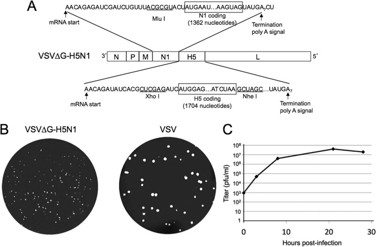
FIG 1.
The VSVΔG recombinant encoding the influenza virus HA and NA genes and representative plaque morphology. (A) Diagram of the VSV recombinant with the insertion of the influenza virus N1 NA and H5 HA genes between the VSV M and L genes in a VSVΔG vector. (B) Plaque morphology of VSVΔG-H5N1 and VSV 2 days after infection of BHK-21 cells. (C) One-step growth curve of VSVΔG-H5N1. BHK-21 cells were infected at time zero with VSVΔG-H5N1 at an MOI of 5, the inoculum was removed after 30 min, cells were washed twice with PBS, and samples of the medium were taken immediately (time zero) and at the indicated times thereafter for subsequent plaque titration.

