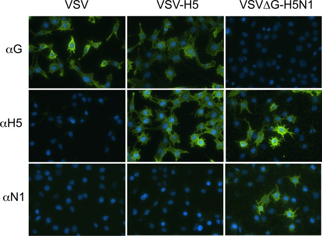
FIG 2.
Indirect immunofluorescence microscopy of VSVΔG-H5N1- and VSV-H5-infected cells. BHK-21 cells were infected with the indicated viruses for 6 h and then fixed in 3% paraformaldehyde. Cells were then incubated with either anti-VSV G mouse monoclonal antibodies, anti-H5 polyclonal sheep serum, or anti-PR8 NA polyclonal mouse serum, followed by anti-mouse or anti-sheep Alexa Fluor 488-conjugated secondary antibody staining. Nuclei are stained with DAPI. Images were taken with a Nikon Eclipse 80i fluorescence microscope.

