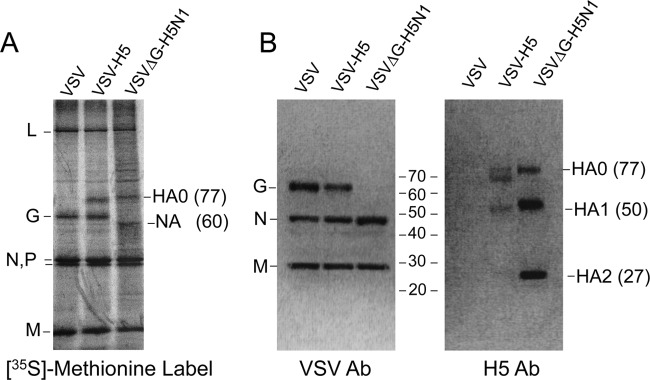
FIG 3.

Metabolic labeling and Western blotting. (A) Metabolic labeling of proteins expressed from recombinant VSV vectors. BHK-21 cells were infected with the indicated viruses and then metabolically labeled with [35S]methionine. Cell lysates were subjected to SDS-PAGE on a 4-to-12% gradient gel, and the protein bands on the dried gel were imaged. Positions of VSV proteins are indicated on the left side of the gel image, and influenza virus proteins are indicated on the right side. (B) Western blot analysis of BHK-21 whole-cell extracts infected with the indicated viruses. Sample lysates were analyzed by SDS-PAGE on a 4-to-12% gradient gel, and Western blotting was performed by using a polyclonal VSV (Indiana serotype) antiserum and the polyclonal H5 HA-specific antibody (Ab) NR-665. The positions of full-length (HA0) and cleaved (HA1 and HA2) H5 HA isoforms are indicated on the right side of the anti-H5 blot.
