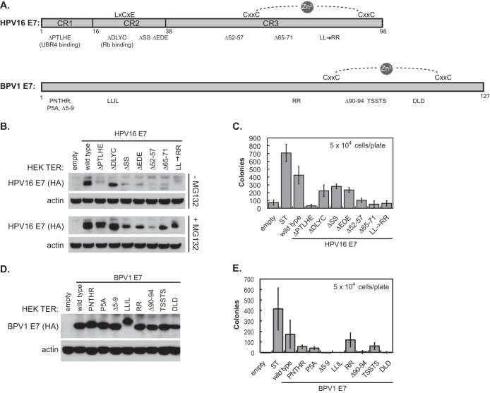
FIG 3.
Regions of HPV16 E7 that contribute to HEK TER anchorage independence. (A) Schematic of HPV16 E7 and BPV1 E7 amino acid deletion and substitution mutants used in the study. The HPV16 E7 schematic was adapted from reference 39. (B) Western blot of stable HPV16 E7 expression in HEK TER cell lines. HPV16 E7 proteins tagged with the hemagglutinin epitope at the C terminus were detected with anti-HA antibody. +MG132, cells were treated with 30 μM the proteasome inhibitor MG132 for 4 h prior to harvest. (C) HEK TER-HPV16 E7 mutant cell lines were plated in soft agar at a density of 5 × 10⁴ cells per 6-cm dish. Cells were plated in triplicate for each condition and incubated at 37°C. Colonies were counted 3 weeks after plating, and values shown in the graphs represent the averages from three independent experiments ± standard deviations. (D) Western blot of stable BPV1 E7 expression in HEK TER cell lines. BPV1 E7 proteins tagged with the hemagglutinin epitope at the C terminus were detected with anti-HA antibody. (E) HEK TER-BPV1 E7 cell lines were plated in soft agar at a density of 5 × 10⁴ cells per 6-cm dish. Cells were plated in triplicate for each condition and incubated at 37°C. Colonies were counted 3 weeks after plating, and values shown in the graphs represent the averages from three independent experiments ± standard deviations.

