FIG 4.
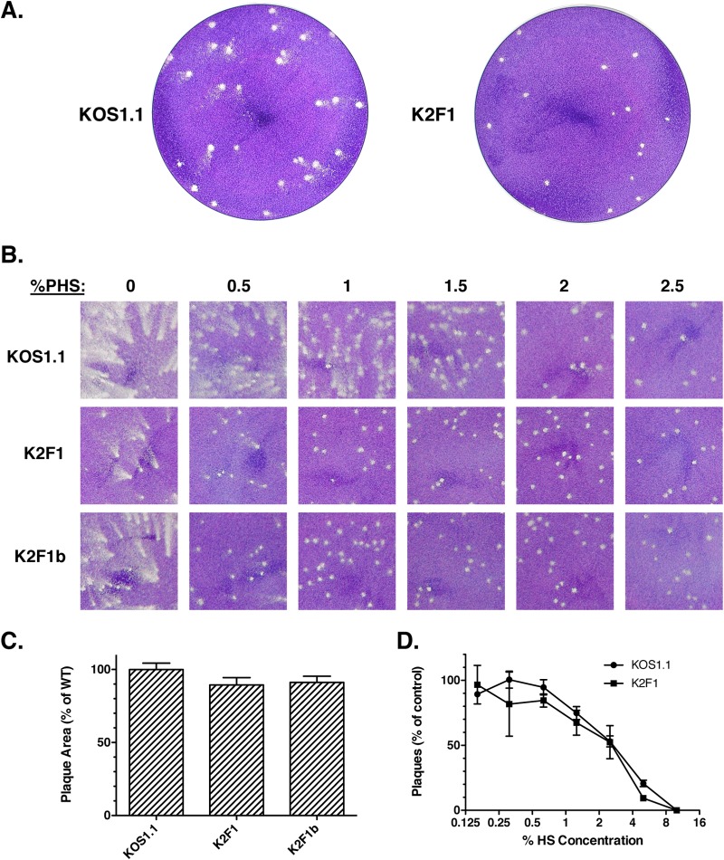
K2F1 forms plaques with an altered morphology. (A) Viral plaques were allowed to form on Vero cells under a liquid medium overlay containing 1% PHS. Plaques were fixed and stained with Giemsa stain at 3 days p.i. (B). K2F1 plaque phenotype depends on the concentration of PHS in the overlay. Equal numbers of PFU of the viral strains shown were subjected to plaque assays on Vero cells. The overlay medium contained differing concentrations of PHS (0 to 2.5%). Plaques were fixed and stained 3 days postinfection. (C) Cell-to-cell spread. Viral stocks were allowed to form plaques in the presence of 10% PHS to completely prevent extracellular spread. Plaques were fixed and stained 3 days postinfection. Digital images were obtained and analyzed by using ImageJ to determine the mean plaque area. Thirty plaques were measured for each virus. (D) K2F1 resembles WT HSV-1 in its sensitivity to neutralization by PHS. Triplicate aliquots containing 200 PFU of KOS1.1 or K2F1 were incubated for 30 min in medium lacking PHS (control) or in medium containing various concentrations of PHS. Following incubation, the virus samples were subjected to a standard plaque assay. Error bars represent standard errors of the means.

