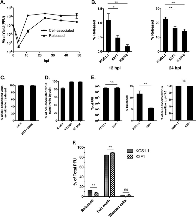
FIG 5.
K2F1 is deficient in release of virus to the extracellular medium. (A) Time course of WT HSV-1 extracellular release. Vero cells were infected with KOS1.1 at an MOI of 5 PFU/cell. At various times after infection, the supernatant and cells were harvested separately and titers were determined for the virus yields in each fraction. (B) K2F1 is deficient in extracellular release. Vero cells were infected in quadruplicate with KOS1.1, K2F1, or K2F1b at an MOI of 5 PFU/cell. At 12 and 24 h p.i., supernatant and cells were harvested separately and the percentage of the total yield that was released into the medium was determined. (C) Most cell-associated HSV-1 progeny are sensitive to pH 3.0 inactivation. Cultures of Vero cells were infected in duplicate at an MOI of 5 PFU/cell, and cells were incubated for 24 h. Cells were harvested by scraping into PBS and low-speed centrifugation. The scraped cells were either incubated for 2 min in PBS as a control or in pH 3.0 buffer to inactivate extracellular virions. After repelleting and suspension in fresh medium, samples were frozen and titers were determined. Prior to titration, one set of low-pH-treated samples was treated by pulsed microsonication to further release intracellular virus. The values represent the percentage of cell progeny that were inactivated by the treatment relative to the PBS control. (D) Most cell-associated HSV-1 progeny are sensitive to trypsin inactivation. Cultures of Vero cells were infected in triplicate and harvested as described for panel C. Cells were resuspended in either PBS, as a control, or in 0.05% trypsin–EDTA, and the resuspended cells were incubated at 37°C for up to 15 min. After repelleting and suspension in fresh medium, samples were frozen and titers were determined. The values represent the percentage of cell-associated progeny that were inactivated by the trypsin treatment relative to the 15-min PBS control. (E) K2F1 virions efficiently reach the cell surface. Vero cells were infected in quadruplicate with KOS1.1 or K2F1 at an MOI of 1 PFU/cell, and the infections were incubated for 20 h. After taking aliquots of the medium to determine release (middle), the scraped cells were treated for 2 min with PBS or pH 3.0 buffer as for panel C to determine sensitivity to low-pH treatment (right). The total viral yields are also shown (left). (F) K2F1 has a higher percentage of progeny virions on the cell surface than does WT HSV-1. Cells were infected in quadruplicate with KOS1.1 or K2F1 at an MOI of 5 PFU/cell and harvested at 24 h p.i. Aliquots of the medium were taken to determine release. The cells were then collecting by scraping and low-speed centrifugation and then extracted twice with TNE buffer. The combined salt washes and the washed cell fractions were frozen, and titers of all fractions were determined. Statistical analyses for results shown in panels B, E, and F were performed using the Student t test; error bars denote standard errors of the means. *, P < 0.05; **, P < 0.01; ns, not significant (P > 0.05).

