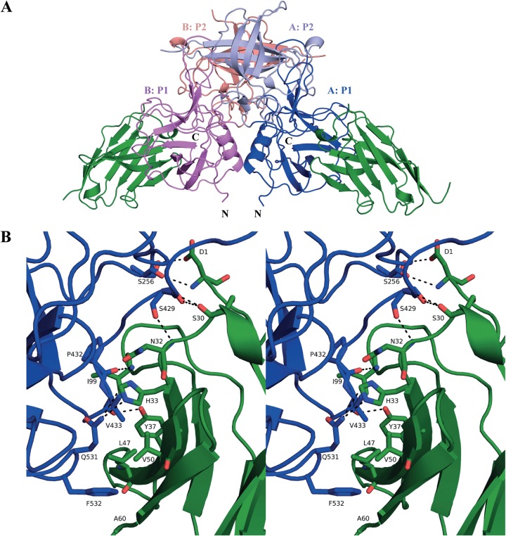
FIG 5.
Structure of GII.10 domain Nano-25 complex. (A) The X-ray crystal structure of the GII.10 domain Nano-25 complex was determined to 1.70-Å resolution. The complex was colored according to GII.10 P domain monomers (chains A and B) and P1 and P2 subdomains, i.e., chain A, P1 (blue); chain A, P2 (light blue); chain B, P1 (violet); chain B, P2 (salmon); and Nano-25 (green). Nano-25 bound to the lower region of the P1 subdomain and involved a monomeric interaction. (B) A closeup stereo view of (chain A) GII.10 P domain- and Nano-25-interacting residues. The GII.10 P domain hydrogen bond interactions involved both side-chain and main-chain interactions (bond length of 2.8 to 3.3 Å), one side chain and one main chain of S256, two main chains and one side chain of S429, one main chain of P432, one main chain of V433, and one side chain of Q531. GII.10 P domain hydrophobic interactions (3.6 to 5.1 Å) involved P432, V433, and F532.

