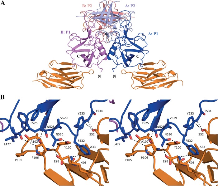
FIG 7.
Structure of GII.10 domain Nano-85 complex. (A) The X-ray crystal structure of the GII.10 domain Nano-85 complex was determined to 2.11-Å resolution. The complex was colored according to Fig. 2, with the exception of Nano-85 (orange). Nano-85 bound to the lower region of the P1 subdomain and involved a monomeric interaction. (B) A closeup stereo view of (chain A) GII.10 P domain- and Nano-85-interacting residues. The P domain hydrogen bond interactions included side-chain and main-chain interactions, one main chain of W528, two side chains of N530, and one main chain of T534. Two additional P domain interactions with Nano-85 were observed, F532, forming a hydrogen bond/electrostatic interaction, and Y533, forming a π donor hydrogen bond. P domain hydrophobic interactions involved L477, F525, V529, and F532.

