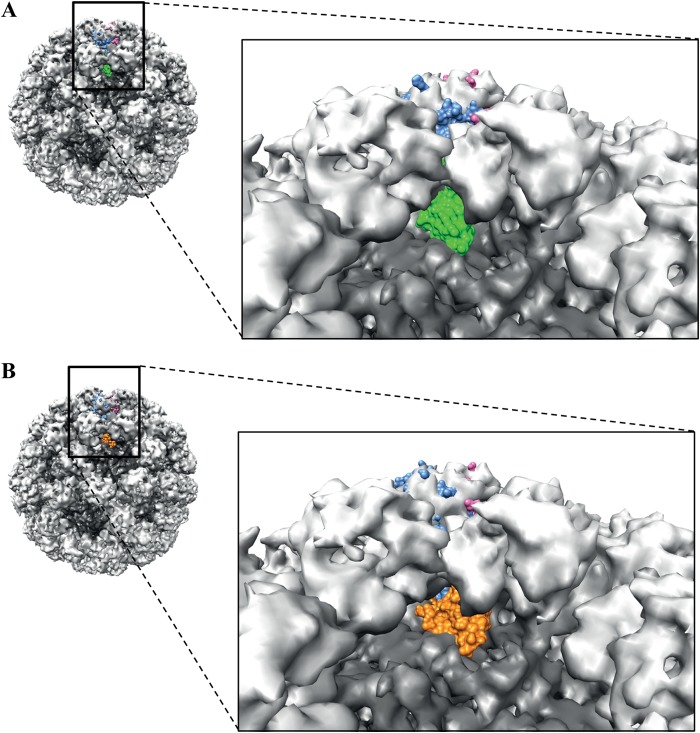
FIG 8.
X-ray crystal structure of the GII.10 P domain Nano-25 complex and GII.10 P domain Nano-85 complex fitted into the cryo-EM GII.10 VLP structure. The cryo-EM VLP structure was colored according to S (dark gray) and P domains (light gray). The P dimer (light blue and pink) from the P domain nanobody complexes was manually fitted into the P dimer on the VLP. (A) The P domain Nano-25 complex fitted on the VLP, showing the position of Nano-25 (green) in the context of the particle. The boxed region shows a close-up view of the complex. Nano-25 clashed with neighboring P domains on the VLP and rested on top of the S domain. (B) The P domain Nano-85 complex fitted on the VLP, showing the position of Nano-85 (orange) in the context of the particle. The boxed region shows a close-up view of the complex. Nano-85 clashed with the neighboring P domains on the particle and rested on top of the S domain.

