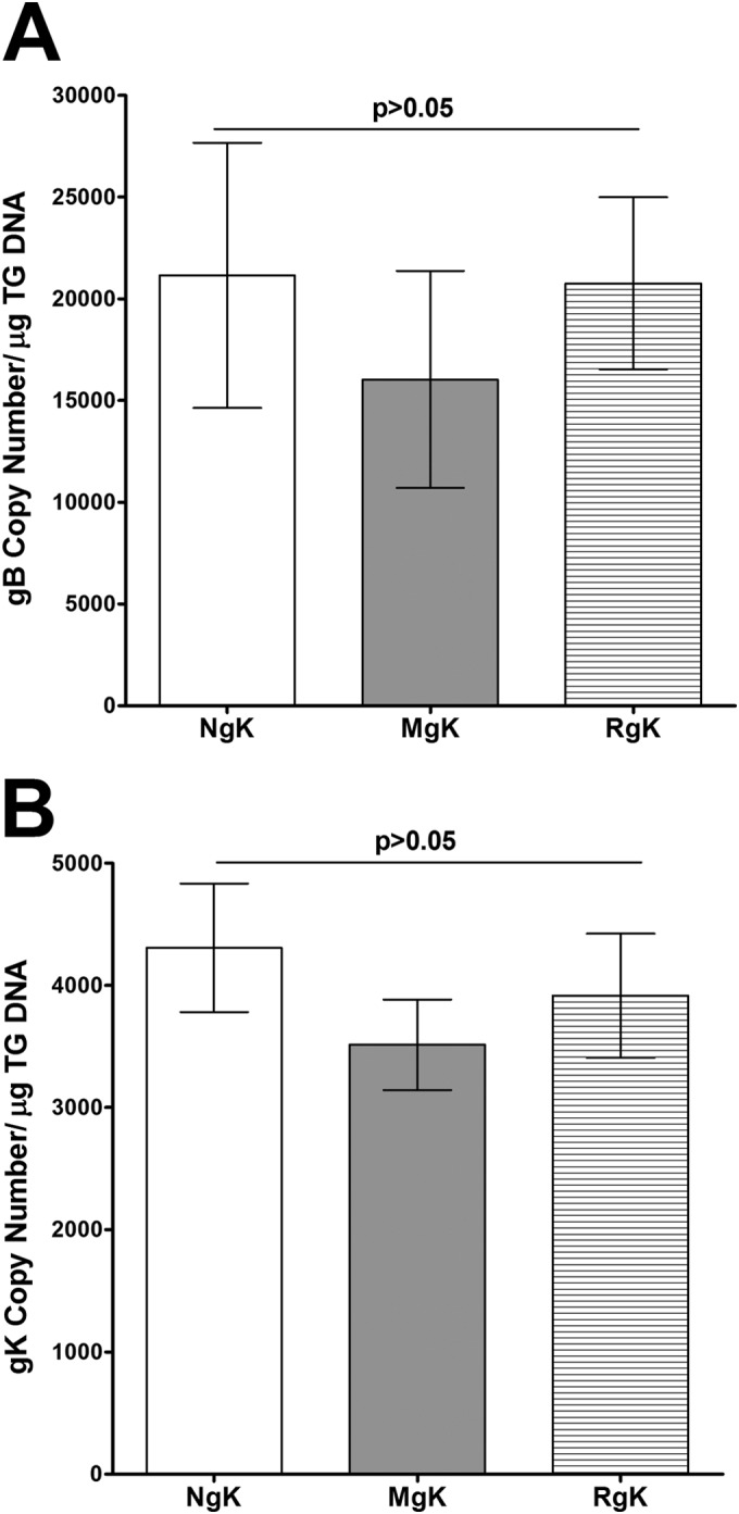
FIG 10.

Detection of gK and gB DNA in TG of latently infected mice. C57BL/6 mice were ocularly infected with 2 × 10⁵ PFU of NgK, MgK, or RgK virus/eye. TG from individual mice were isolated 28 days p.i., and TaqMan qPCR was performed as described in Materials and Methods. GAPDH was used as an endogenous control to normalize the relative expression of gK and gB DNA in infected TG. The relative copy numbers of the HSV-1 gB and gK genes were calculated using standard curves as described in Fig. 9. Each point represents the mean ± the SEM from 12 TG (six mice). (A) gB DNA; (B) gK DNA.
