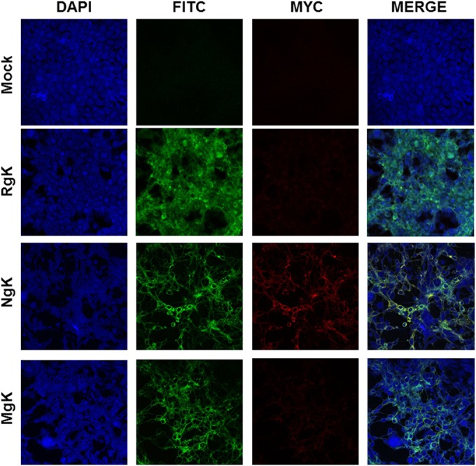
FIG 6.

Immunostaining of NgK- or MgK-infected cells in vitro. RS cells were infected with the NgK, MgK, or RgK at an MOI of 5 PFU/cell for 24 h or were mock infected. At 24 h p.i., the cells were fixed, permeabilized, blocked, and stained with anti-HSV-1 gC (green), anti-myc (red), and DAPI nuclear stain (blue). The slides were fixed, and photomicrographs are shown at ×40 direct magnification. Colocalization is visualized as yellow.
