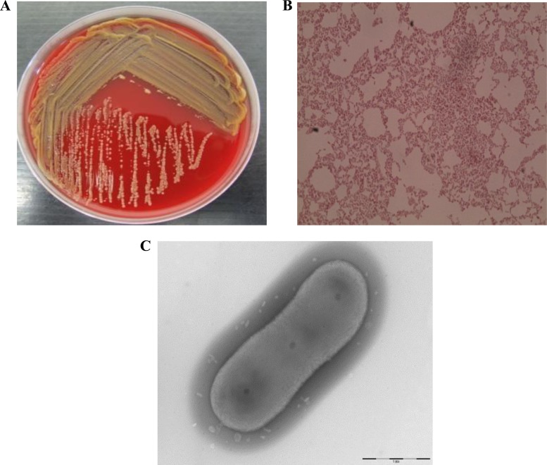
FIG 1.
(A) Chryseobacterium oranimense G311 yellow isolate on Columbia agar with 5% sheep blood (bioMérieux) at 37°C; (B) Gram staining image of Chryseobacterium oranimense G311 viewed at ×100 magnification; (C) transmission electron microscopic image of Chryseobacterium oranimense G311 using a Morgani 268D TEM (Philips) at an operating voltage of 60 kV.

