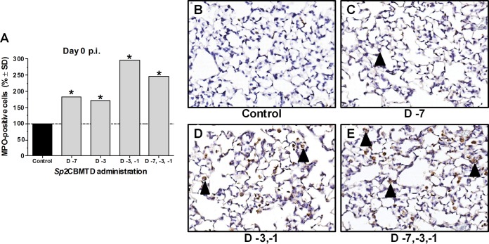
FIG 5.
Infiltration of mouse lung tissues with neutrophils after administration of Sp2CBMTD. Sp2CBMTD was administered as described in the Fig. 2 legend. Mouse lungs were obtained on day 0 p.i., fixed in 10% neutral-buffered formalin, and stained with myeloperoxidase (MPO), a specific marker for neutrophils. The MPO-stained sections were blinded for pathology evaluation. The presence of antigens was quantified by capturing digital images of whole-lung sections using an Aperio ScanScope XT slide scanner (Aperio Technologies) and then manually outlining entire fields together with areas of noticeably decreased or absent MPO staining. The percentage of the lung field with reduced staining coverage was calculated using the Aperio ImageScope software and expressed as a relative value over virus-infected PBS-treated (control) animals (A). Representative MPO-stained lung images of the control animals (B) and animals given single-dose (C), double-dose (D), or triple-dose (E) regimens of Sp2CBMTD before H7N9 virus infection. Magnification, ×20. The black arrowhead indicates neutrophils. *, P < 0.0001 compared with results for the control group, one-way ANOVA.

