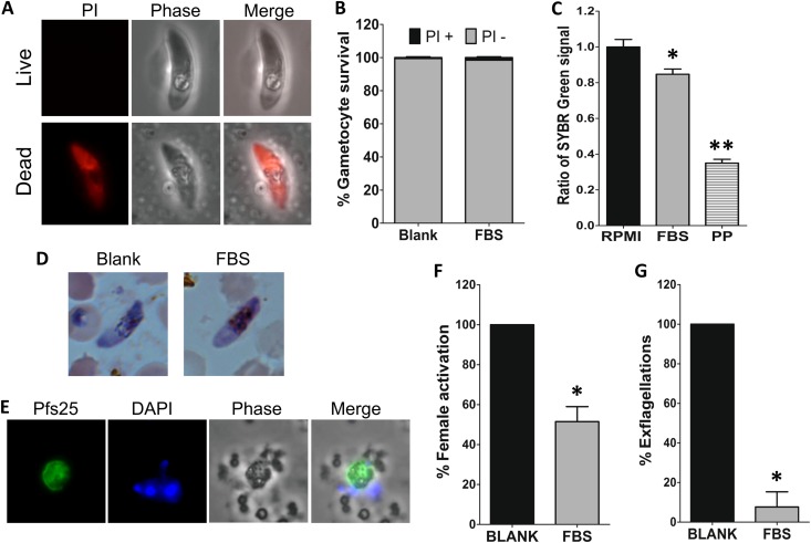
FIG 4.
FBS0701 inhibits P. falciparum gametocyte activation. (A) Propidium iodide staining of live (top) or dead (bottom) gametocytes. (B) Quantification of propidium iodide staining of gametocytes incubated in the absence or presence of 25 μM FBS0701 (FBS) for 48 h (P > 0.05, Fisher's exact two-tailed test). (C) Relative fluorescence of SYBR green I/CyQUANT staining of gametocytes incubated in the presence of RPMI, 25 μM FBS0701, or 10 μM pyrvinium pamoate (PP) for 48 h. The values were normalized to the RPMI control. Statistical significance was determined by one-way ANOVA with the Bonferroni multiple-comparison test (*, P < 0.05; **, P < 0.001). (D) Light microscopy of Giemsa-stained gametocytes. (E) Immunofluorescence detection of Pfs25 protein on P. falciparum female gametes after 4 h of incubation in gametocyte activation medium. DAPI, 4′,6-diamidino-2-phenylindole. (F) Percent female gametocyte activation determined by Pfs25 detection in the presence or absence of 25 μM FBS0701 for 48 h. The results were normalized to the blank control. Statistical significance was determined by Student's t test (*, P = 0.0008). (G) Percent male gametocyte exflagellation after incubation with 25 μM FBS0701 for 48 h. The results were normalized to the blank control. The error bars represent the standard error of the mean. Statistical significance was determined by Student's t test (*, P < 0.0001).

