FIGURE 1:
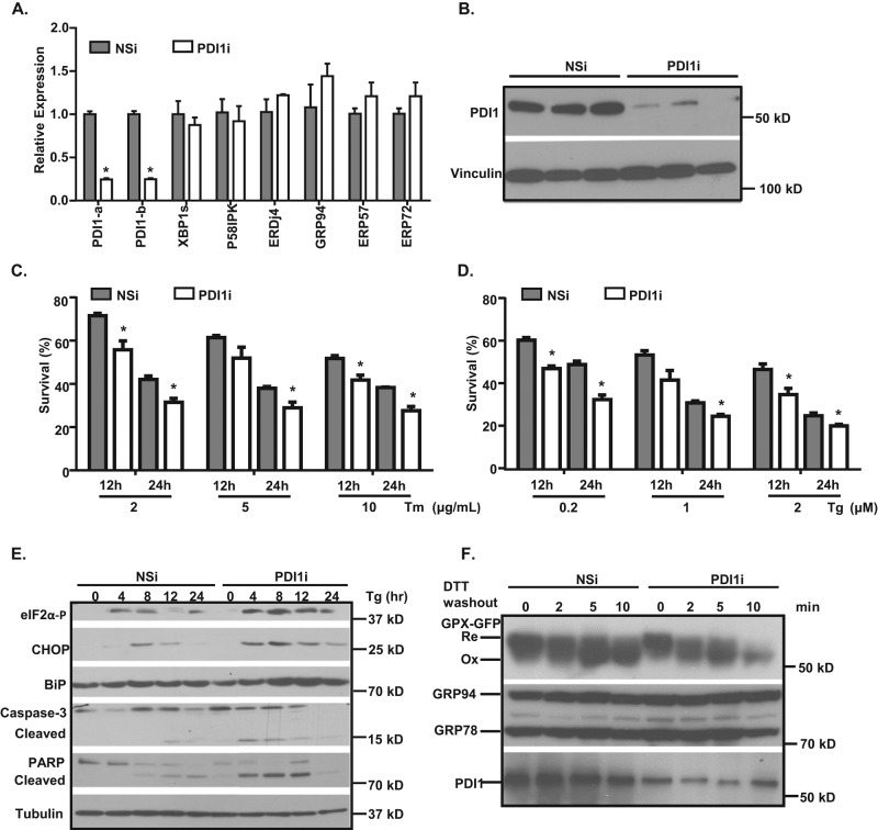
Knockdown of Pdi1 sensitizes cells to ER stress. (A, B) Adenoviral expression of Pdi1 shRNA knocks down PDI1 levels in McA cells. (A) Relative abundance of mRNA expressed in control (NSi) and Pdi1-knockdown (PDI1i) cells. mRNA levels were normalized to actin levels. Values are relative to mRNA levels of control cells. *p < 0.01 vs. NSi. (B) PDI1 protein levels in control (NSi) and Pdi1-knockdown (PDI1i) cells. Cell lysates were subjected to immunoblotting analyses using PDI1 and vinculin antibodies. (C–E) Pdi1-knockdown cells are more sensitive to ER stress. (C, D) Cell survival was measured by cell counting after treatment with different concentration of Tm or Tg. *p < 0.01 vs. NSi in the same treatment group. (E) Protein levels of ER stress markers in control (NSi) and Pdi1-knockdown (PDI1i) cells. Cells were treated with 0.2 μM Tg for indicated time periods, and cell lysates were immunoblotted for cleaved caspase 3, PARP, eIF2α-P, CHOP, and BiP. (F) ER redox balance is not altered in Pdi1-knockdown cells. Cells expressing Grx-roGFP-iEER were treated with 10 mM DTT for 10 min and washed out. The oxidation status of Grx-roGFP-iEER was analyzed by nonreducing SDS–PAGE and immunoblotting with GFP, KDEL (GRP78 and GRP94), and PDI antibodies.

