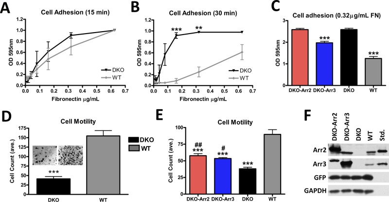
FIGURE 2:
Arrestins regulate cell migration and adhesion. (A, B) Adhesion was measured by plating cells on serial dilutions of FN (0.01–1.25 μg/ml) for 15 (A) or 30 (B) min. The data were analyzed by one-way analysis of variance (ANOVA) with arrestin type as the main factor, which was highly significant at 30 min. DKO cells showed a dramatic increase in their ability to adhere compared with WT cells, ***p < 0.001, **p < 0.01. Means ± SD from three experiments. (C) Adhesion of DKO cells expressing arrestin-2 + GFP, arrestin-3 + GFP, or GFP alone (controls). Cells were plated on 0.32 μg/ml FN. Means ± SD from 24 data points in three experiments. ***p < 0.001 compared with DKO. (D) Cells were plated in Transwell chambers coated with 0.32 μg/ml FN and allowed to migrate for 4 h. Cells were counted in six fields/chamber in each of four independent experiments. The data were analyzed by one-way ANOVA with cell type as the main factor, ***p <0.001. Insets, representative membranes postmigration. (E) Migration of DKO cells expressing arrestin-2 and GFP or arrestin-3 and GFP, or cells expressing GFP only (DKO and WT). Means ± SD from 5 fields/chamber from three independent experiments performed in duplicate analyzed by one-way ANOVA with cell type as the main factor. ***p < 0.001 compared with WT. DKO-Arr2, ##p < 0.01, and DKO-Arr3, #p < 0.05, compared with DKO. (F) Arrestin expression in DKO cells was determined using arrestin-2– or arrestin-3–specific antibodies, with corresponding purified bovine arrestins (0.1 ng/lane) run as standards.

