FIGURE 3:
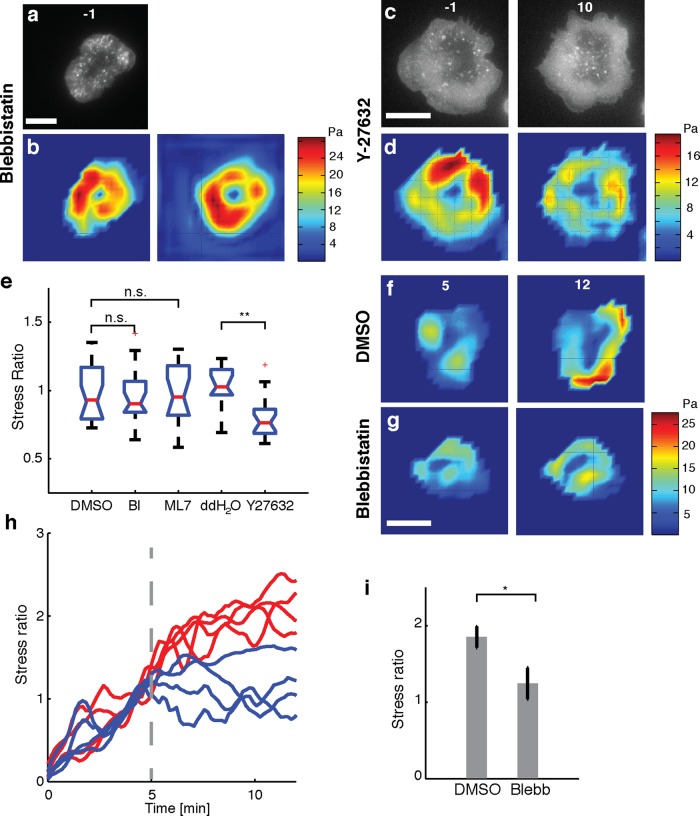
Effect of myosin II activity on cellular force generation. (a) Fluorescence image of an EGFP-actin Jurkat T-cell on an elastic substrate 1 min before addition of 50 μM blebbistatin. (b) Traction stress color map of the same cell 1 min before (left) and 9 min after (right) addition of blebbistatin. (c) Fluorescence images of an EGFP-actin Jurkat T-cell on an elastic substrate 1 min before (left) and 9 min after (right) application of 100 μM Y-27632. (d) Traction stress color maps of the same cell before and after addition of Y-27632. (e) Comparison of the after-to-before ratios of traction stresses upon addition of blebbistatin (N = 20 cells) and ML7 (N = 17 cells) with control (DMSO carrier) and comparison of traction stress ratios upon addition of Y-27632 (N = 20 cells) with double-distilled H2O control (N = 11 cells). The average stresses in a 3- min time interval just before addition of drug and in the time interval 9–12 min after addition of drug were used to compute the ratios. **p < 0.01. (f, g) Traction stress color maps for example cells (at the indicated time points after stimulation). Drug or vehicle was added at 5 min after stimulation (f, DMSO; g, blebbistatin). (h) Traces of the total force exerted by four example cells with drug addition 5 min after stimulation (vertical dashed line). The total force is normalized to the value exerted at 5 min after stimulation. Red lines indicate vehicle, and blue lines indicate the time of blebbistatin addition. (i) Summary statistics of the stress ratio after drug addition for cells averaged between 9 and 12 min after stimulation. N = 13 for blebbistatin and N = 12 for DMSO (p < 0.05, Wilcoxon's rank sum test). Scale bars, 10 μm.

