FIGURE 1:
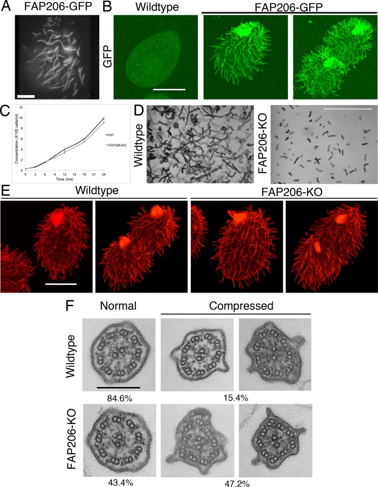
FAP206 localizes to the ciliary axoneme, and knockout of FAP206 results in cilia-related defects. (A) TIRF image of a live cell expressing FAP206-GFP under the native promoter. (B) A wild-type (negative control) cell (left) and a cell expressing FAP206-GFP under the native promoter (right) extracted with Triton X-100, fixed with paraformaldehyde, and imaged for GFP using a confocal microscope. (C) Culture growth rates for a wild-type CU428 and an FAP206-KO strain. Each data point represents an average for three independent experiments. (D) Paths of swimming wild-type and FAP206-KO cells recorded for 1 s. The average swim velocities were 170 μm/s for the wild type and 50 μm/s for FAP206-KO. (E) Immunofluorescence images of tubulin for wild-type and FAP206-KO cells. For each genotype, an interphase (left) and a dividing cell (right) are shown. (F) Classical TEM images of cilia cross-sections that are either circular (left) or compressed (right). The percentages represent fractions of either circular or compressed axonemes (n = 52 for wild type, n = 48 for FAP206-KO). Scale bars, 20 μm (A, B, and E), 1 mm (D), 0.2 μm (F).

