FIGURE 2:
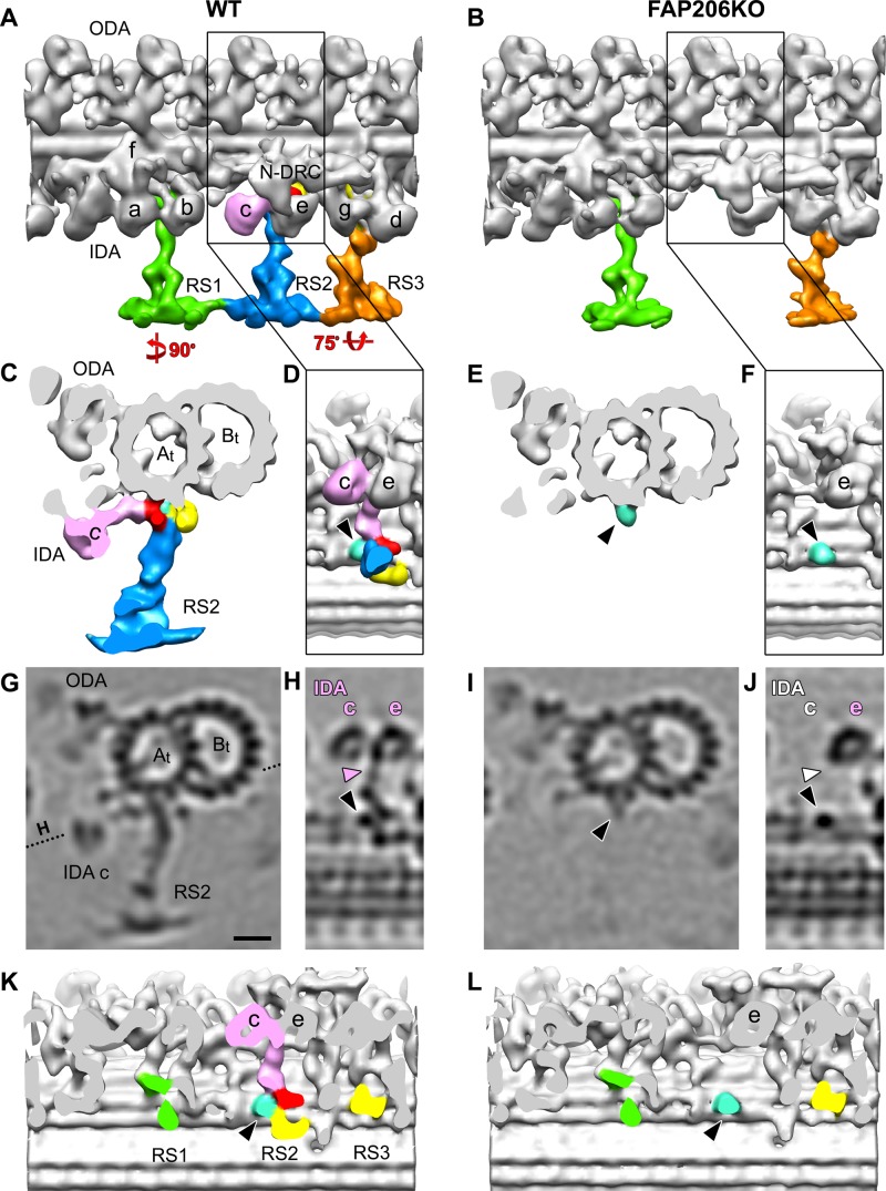
Deletion of FAP206 leads to loss of RS2 and associated dynein c in the 96-nm repeat. Isosurface renderings (A–F, K, L) and tomographic slices (G–J) show the averaged 96-nm axonemal repeats of wild type (A, C, D, G, H, K) and FAP206-KO (B, E, F, I, J, L) in longitudinal (A, B, D, F, H, J–L), cross-sectional (C, E, G, I), and bottom views looking from the central pair toward the doublet microtubule (D, F, H, J–L); the dotted line in G indicates the orientation of the tomographic slice shown in H and J. There are three radial spokes: RS1 (green), RS2 (blue), and RS3 (orange) in wild type (A), whereas RS2 is missing in FAP206-KO (B, E, I). The RS2 base is composed of three regions: front (red), back (yellow), and side prongs (light blue and/or black arrowheads). These three regions are connected with each other and form the attachment of RS2 to the doublet A-tubule (At). The back prong of RS2 and the RS3 base (yellow) were previously identified as parts of the CSC (Heuser et al., 2012b). IDA c (pink), which anchors with its tail (pink arrowhead in H) to the A-tubule through the front prong of the RS2 base in WT, is also missing in FAP206-KO (white labels in J, where the IDA c with tail should be). (K, L) Densities representing radial spoke heads and stems were removed to visualize the microtubule-attachment sites of the spoke bases. Note that in the FAP206-KO mutant, the main difference from wild type is the complete absence of the RS2 front prong (red), RS2 back prong (yellow), and dynein c; in contrast, RS1, RS3, and the RS2 side prong (light blue, black arrowhead) appear unaffected in FAP206-KO. All structures shown are subtomogram averages without prior classification analysis. Scale bar, 10 nm.

