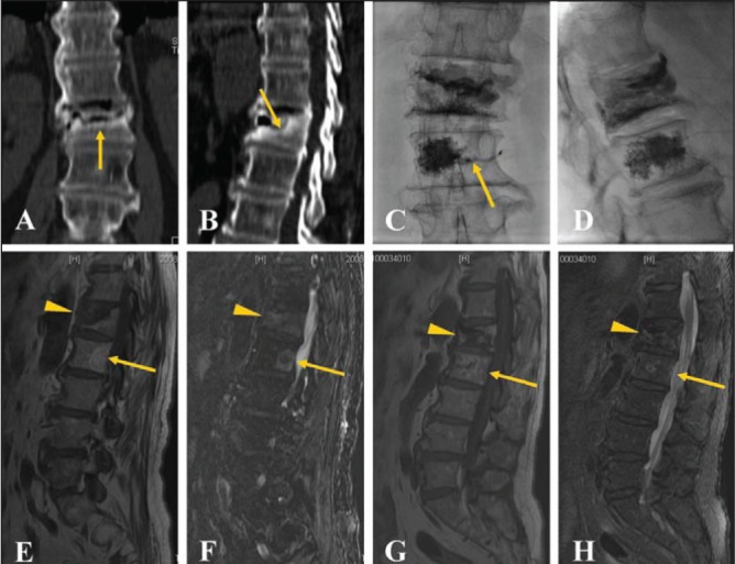
Figure 1).

A 74-year-old male patient with persistent back pain and a visual analogue scale score of 7 for >10 months. Coronary (A) and sagittal (B) computed tomography reconstruction demonstrates a border of osteosclerosis at the fracture site (arrow). Anteroposterior (C) and lateral (D) plain films show bone cement injected into the L1 and L2 vertebral bodies with slight vein leakage (arrow) at the L2 level. Magnetic resonance imaging reveals low signal (arrowhead) on T1WI images (E) and high signal (arrowhead) on T2WI (F) at the L1 level before percutaneous vertebroplasty. Note also a hemangioma (arrow) at the L2 level. Magnetic resonance imaging displays low signal (arrowhead) on T1WI (G) and slightly high signal (arrowhead) on T2WI (H) images at the L1 level one year after percutaneous vertebroplasty with stability of the vertebral body without obvious focal kyphosis
