
Figure 1.
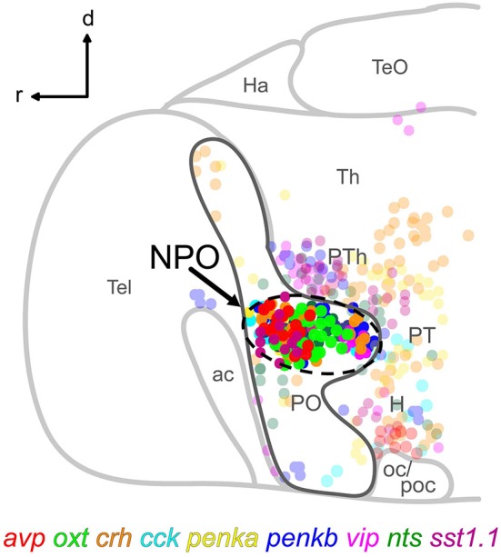
Schematic lateral view of a 5 dpf larval zebrafish brain showing the location of the NPO (dashed line) within the otpa-positive part (dark gray line) of the preoptic area, and the spatial distribution of nine cell types expressing the indicated neuropeptides. Cells clustering within the NPO are opaque. For more details, the reader is referred to our previously reported chemoarchitectural map (Herget et al., 2014). Abbreviations: ac, anterior commissure; d, dorsal; H, hypothalamus; Ha, habenula; NPO, neurosecretory preoptic area; oc, optic chiasm; PO, preoptic area; poc, postoptic commissure; PT, posterior tuberculum; PTh, prethalamus; r, rostral; Tel, telencephalon; TeO, optic tectum; Th, thalamus.